New X-Ray Contrast Agent Improves Soft Tissue Imaging with Lower Dose

Key Takeaways
- A new positively charged contrast agent developed at Northeastern University is changing how cartilage is imaged on X-rays. Unlike conventional iodine-based agents, this new agent is engineered with a net positive charge that creates electrostatic attraction to the negatively charged proteoglycans in cartilage.
- The charge-based mechanism turns the agent into a functional indicator of cartilage health. Its differential uptake creates a visible contrast gradient on X-ray, meaning regions of early degeneration that would be invisible on conventional arthrography become detectable for the first time.
- The agent requires up to 40 times less contrast volume than conventional methods. This reduction makes high-resolution joint imaging safer and more accessible, particularly for patients with chronic kidney disease, who have historically been at elevated risk for contrast-induced nephropathy from iodine-based agents.
- The technology is still in preclinical research and has not yet entered human trials. The path to clinical use will involve large animal studies, full toxicology profiling, an FDA Investigational New Drug review, and three phases of human trials, a process that typically takes several years even for agents with strong early data.
- As contrast imaging evolves, so does the need for reliable supervision. At ContrastConnect, we provide healthcare facilities with virtual contrast supervision delivered by experienced radiologists through a secure, HIPAA-compliant platform.
A New Contrast Agent Is Changing How Doctors See Cartilage
Researchers at Northeastern University have engineered an X-ray contrast agent that uses positive charge to bind directly to cartilage, producing clearer soft tissue images at roughly 40 times lower volume than standard iodine arthrography. The mechanism is electrostatic: cartilage carries a net negative charge, so the agent is pulled into the tissue and held there for sharp visualization.
For imaging teams, this means reduced risk for patients with chronic kidney disease, faster procedures, and the ability to detect early cartilage degeneration that conventional methods often miss.
The agent remains in preclinical research, but it reflects a larger shift in contrast medicine that demands evolving supervision standards. ContrastConnect helps imaging networks stay ahead with virtual contrast supervision delivered through a secure, HIPAA-compliant platform, keeping facilities compliant and patients safe regardless of which agents enter the workflow.
ContrastConnect: Virtual Contrast Supervision That Never Misses
Built by Radiologists | 75,000+ Monthly Contrast Exams | Trusted Nationwide

Built for Imaging Networks:
- Virtual Contrast Supervision: Radiologists provide immediate CMS-compliant supervision through a secure, HIPAA-compliant platform for outpatient facilities and hospital networks.
- Unmatched Experience: 130+ contrast reactions treated monthly with 3,700+ technologists certified.
The ContrastConnect Difference:
- ✓ Radiologist-owned with superior clinical expertise
- ✓ Always-on platform with guaranteed compliance
- ✓ Audit-ready documentation for CMS reviews
- ✓ Cost-efficient alternative to onsite staffing
Safety & Compliance You Trust:
Helping imaging centers reduce cancellations, extend hours, and scale operations without adding on-site radiologists. Response times measured in seconds.
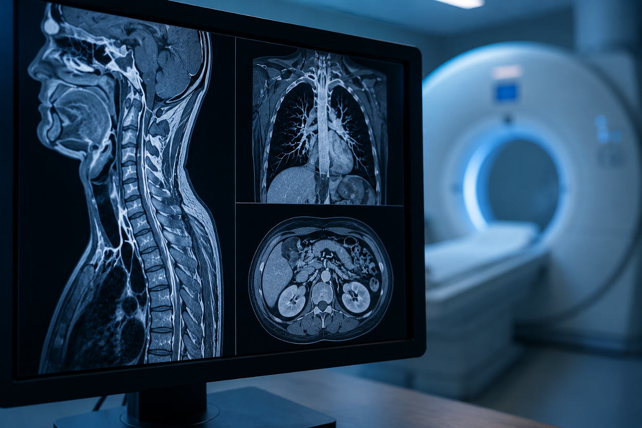
Why X-Rays Struggle with Soft Tissue
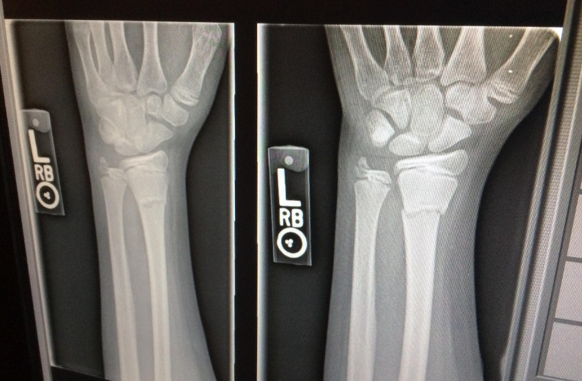
X-rays excel at imaging dense, calcium-rich bone but struggle to image soft tissues such as muscle, cartilage, and tendons. This is a fundamental physics limitation that no equipment upgrade can fully solve. Cartilage is especially problematic: it lacks a significant blood supply and radiographic density, so it disappears into the background on standard X-rays, forcing clinicians to rely on indirect measurements like joint-space narrowing.
Iodine-based contrast agents have been used as a workaround, but they don't concentrate well in cartilage and leak quickly into surrounding tissue. This pushes clinicians to use higher injection volumes that carry real risks, particularly for patients with reduced kidney function. The result is a diagnostic gap that often closes the window for early intervention.
How the New Positively Charged Contrast Agent Works
The core innovation here is electrochemical, not pharmaceutical. The extracellular matrix of cartilage contains proteoglycans that carry a net negative charge throughout the tissue. Conventional iodine contrast agents are either neutral or slightly negative, meaning they have no particular affinity for cartilage and disperse freely once injected.
The new agent developed at Northeastern University is engineered with a net positive charge. When injected into the joint space, electrostatic attraction pulls the agent toward the negatively charged proteoglycan network within the cartilage.
Instead of diffusing away, the contrast accumulates and is held in place, making it available for X-ray detection at much lower concentrations than would otherwise be required.
Why Charge Matters: Attraction to Cartilage
This charge-based mechanism improves imaging and makes the agent a functional indicator of cartilage health. Healthy cartilage has a high density of negatively charged proteoglycans, which means it attracts and retains more of the positively charged contrast.
Degraded cartilage, where proteoglycan content is reduced, attracts less. That differential uptake creates a visible contrast gradient on X-ray images that directly correlates with tissue integrity.
Regions of early degeneration that would be invisible on conventional arthrography become detectable because the contrast agent distributes unevenly based on tissue charge density. This is a meaningful leap in diagnostic specificity for joint imaging.
How the Agent Enters & Leaves the Body
The positively charged contrast agent is administered via intra-articular injection, the same delivery route used in conventional contrast arthrography. Once inside the joint, electrostatic attraction draws the agent into the cartilage matrix, preventing it from dispersing freely.
This targeted accumulation means the agent reaches diagnostically useful concentrations within the tissue at a fraction of the volume required by standard iodine-based methods. Clearance is expected to follow natural joint fluid turnover, with the agent eventually draining into surrounding lymphatic and vascular structures as the charge-based binding weakens over time.
Dose Reduction: 40 Times Less Contrast Required
One of the most clinically significant findings from this research is the dramatic reduction in required contrast volume. Developers noted that imaging with the new positively charged agent can be performed using approximately 40 times less contrast than conventional arthrography methods.
Why Lower Dose Matters for Kidney Disease Patients
Iodine-based contrast agents are filtered through the kidneys, and in patients with pre-existing renal impairment, the contrast load can accelerate kidney damage or trigger contrast-induced nephropathy. This risk has historically forced clinicians to either limit imaging quality, choose alternative modalities like MRI, or accept elevated procedural risk in diagnostically ambiguous cases.
A reduction of this magnitude significantly changes that equation. Even patients with moderate-to-severe chronic kidney disease may fall within a safer exposure window using the new agent, potentially opening up high-resolution joint imaging to a group that has been systematically underserved by conventional arthrography.
How Reduced Volume Improves Safety Profiles
Beyond renal considerations, lower injection volumes reduce mechanical stress on the joint capsule, decrease the risk of procedure-related discomfort, and limit exposure to any adjunct agents used in the injection preparation.
Smaller volumes also mean faster administration and potentially shorter post-procedure observation windows, which are practical advantages in high-throughput imaging environments.
Where the Research Stands Right Now
The positively charged contrast agent has demonstrated compelling results in preclinical studies, but it has not yet entered human clinical trials.
The path from laboratory research to routine clinical use involves multiple regulatory and safety hurdles that take years to clear, even for agents with strong early data.
That said, the preclinical results are substantive enough to warrant serious attention from the imaging and orthopedic communities. The research comes from Northeastern University's bioengineering program, and the findings have been published in peer-reviewed scientific journals, including ACS Nano.

Mouse Studies & What They Showed
Preclinical testing in mouse models demonstrated that the positively charged contrast agent accumulated selectively in cartilage tissue following intra-articular injection. The agent produced clear X-ray visualization of soft-tissue structures that would be invisible on standard radiographs, and did so at dramatically lower concentrations than conventional iodine-based agents.
Critically, the differential uptake between healthy and degraded cartilage was measurable, meaning the imaging result carried diagnostic information about tissue composition, not just tissue location. The animal studies also provided early safety data, showing no significant adverse tissue responses at the doses tested.
The Road to Human Clinical Trials
Translating a preclinical contrast agent into an approved clinical tool requires moving through a multi-phase regulatory process. Before human trials can begin, the agent must meet a series of escalating safety and efficacy benchmarks, including larger-animal studies, toxicology assessments, and an investigational new drug application reviewed by the FDA. Each phase builds the evidence base required to justify first-in-human dosing.
The timeline for this process varies considerably depending on funding, institutional support, and whether the agent qualifies for any expedited review pathways. Key milestones the research team will need to hit include:
- Completion of large animal model studies with reproducible imaging and safety data.
- Full toxicology and pharmacokinetics profiling across multiple dose ranges.
- Investigational New Drug (IND) application submission and FDA review.
- Phase I human trials focused on safety and preliminary dosing in healthy volunteers.
- Phase II trials assessing diagnostic efficacy in patients with joint degeneration.
- Phase III comparative trials against the current standard-of-care arthrography methods.
Get Safe Remote Contrast Supervision at ContrastConnect

As contrast imaging technology advances, the protocols and oversight surrounding contrast administration must keep pace. When you are working with established iodine-based agents or preparing for next-generation options, having reliable remote supervision infrastructure in place is key for patient safety and regulatory compliance.
At ContrastConnect, we provide healthcare professionals with expert remote contrast supervision services. We help imaging teams meet safety standards, manage adverse event protocols, and maintain oversight across facilities where on-site physician supervision may not always be available. Start your coverage assessment to bring reliable, always-on supervision to your imaging team.
Start Your Coverage Assessment →
Frequently Asked Questions (FAQs)
What is a contrast agent used for in X-ray imaging?
A contrast agent is a substance injected into the body to make specific tissues or structures more visible on X-ray images. In joint imaging, contrast agents are injected directly into the joint space to improve visualization of soft tissue structures, such as cartilage, ligaments, and the joint capsule, which would otherwise be nearly invisible on standard radiographs.
Why is soft tissue hard to see on a standard X-ray?
Soft tissue is difficult to visualize on a standard X-ray because it does not absorb X-rays efficiently. X-ray imaging works by measuring differential absorption. Dense structures like bone absorb a large portion of radiation and appear bright white, while soft tissues like cartilage, muscle, and fat allow most radiation to pass through, rendering them faint or invisible.
How is the new contrast agent different from iodine-based agents?
Conventional iodine-based contrast agents are electrically neutral or slightly negative, meaning they have no chemical affinity for cartilage tissue. The new positively charged contrast agent developed at Northeastern University is electrostatically attracted to the negatively charged proteoglycan molecules that make up cartilage's extracellular matrix.
This targeted accumulation allows the agent to concentrate directly within cartilage tissue at roughly 40 times lower volume than conventional methods, while also producing a differential uptake pattern that reflects cartilage composition and health.
Is the new contrast agent safe for patients with kidney disease?
Based on the current preclinical data, the dramatically reduced volume requirement is a meaningful safety advantage for patients with renal impairment. Iodine-based contrast agents are renally cleared, and higher volumes increase the risk of contrast-induced nephropathy.
Because the new positively charged agent achieves effective imaging at a fraction of the conventional dose, it may fall within a safer exposure threshold for patients who currently cannot safely undergo standard contrast arthrography.
What are the benefits of virtual contrast supervision with ContrastConnect?
At ContrastConnect, we provide imaging facilities with CMS-compliant virtual contrast supervision delivered by experienced radiologists through a secure, HIPAA-compliant platform, closing the gap when on-site physician oversight isn't available.
The benefits include reduced cancellations, extended operating hours, audit-ready documentation for regulatory reviews, and faster response times measured in seconds, all without the cost of full-time on-site staffing.
*Note: Information provided is for general guidance only and does not constitute medical, legal, or financial advice. Pricing estimates and regulatory requirements are current at the time of writing and subject to change. For personalized consultation on imaging center operations and virtual contrast supervision, contact ContrastConnect.
Trusted Nationwide


.avif)


1,000,000
Contrast exams supervised annually
75,000+
Hours of supervision monthly
3,900+
Technologists certified
100s
Of imaging partners nationwide
130+
Contrast reactions treated monthly
100%
Requested hours covered
Connect with us.

