4 Types of Contrast Media in Radiology: Examples & Use Cases

Key Takeaways
- Contrast media enhances visualization of internal structures during radiological imaging, with four primary types used: iodinated, gadolinium-based, barium sulfate, and microbubble agents.
- Each contrast medium serves specific imaging modalities; iodinated agents for X-rays/CT scans, gadolinium for MRI, barium for GI tract visualization, and microbubbles for ultrasound enhancement.
- The administration method (oral, intravenous, or rectal) depends on the area being examined and the type of contrast media used.
- Proper contrast media selection dramatically improves diagnostic accuracy for conditions ranging from vascular abnormalities to inflammatory diseases and tumors.
- ContrastConnect provides CMS-compliant virtual supervision for facilities administering contrast media, with qualified radiologists supervising 75,000+ contrast exam hours monthly, full technologist training, and reliable coverage across imaging networks nationwide.
What Are The 4 Types of Contrast Media in Radiology?
The four primary types of contrast media used in radiology today are iodinated contrast agents, gadolinium-based contrast agents (GBCAs), barium sulfate suspensions, and microbubble agents.
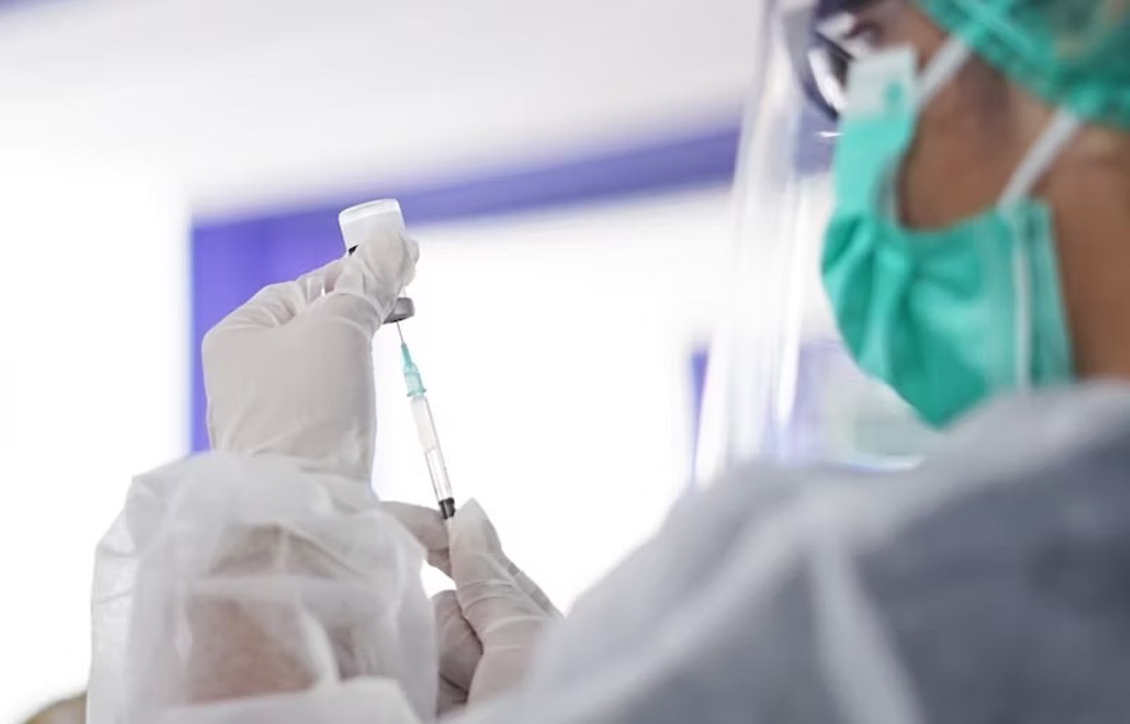
Iodinated contrast agents work by absorbing X-rays through iodine atoms, making them the standard for conventional radiography, fluoroscopy, and CT scans, with non-ionic agents like iohexol (Omnipaque), iopamidol (Isovue), and iodixanol (Visipaque) dominating modern practice due to improved safety profiles.
Gadolinium-based contrast agents work through paramagnetic effects that shorten T1 relaxation time, creating bright signal intensity on MRI T1-weighted images, with macrocyclic agents like gadobutrol (Gadavist) and gadoterate meglumine (Dotarem) offering greater stability than linear agents.
Barium sulfate suspensions remain the leading standard for gastrointestinal imaging through different density formulations (high, medium, low) selected by clinical application, while microbubble contrast agents like Definity, Optison, and Lumason expand ultrasound from anatomical to functional imaging by producing harmonic frequencies under ultrasound waves.
For imaging facilities administering any type of contrast media, ContrastConnect provides virtual contrast supervision by qualified radiologists with experience across 75,000+ monthly contrast exam hours and 130+ reactions treated monthly.
ContrastConnect: Virtual Contrast Supervision That Never Misses
Built by Radiologists | 75,000+ Hours Monthly Contrast Exams | Trusted Nationwide

Built for Imaging Networks:
- Virtual Contrast Supervision: Radiologists provide immediate CMS-compliant supervision through a secure, HIPAA-compliant platform for outpatient facilities and hospital networks.
- Unmatched Experience: 130+ contrast reactions treated monthly with 3,900+ technologists certified.
The ContrastConnect Difference:
- ✓ Radiologist-owned with superior clinical expertise
- ✓ Always-on platform with guaranteed compliance
- ✓ Audit-ready documentation for CMS reviews
- ✓ Cost-efficient alternative to onsite staffing
Safety & Compliance You Trust:
Helping imaging centers reduce cancellations, extend hours, and scale operations without adding on-site radiologists. Response times measured in seconds.
4 Major Types of Contrast Media Used in Radiology Today

1. Iodinated Contrast Media for X-rays & CT Scans
Iodinated contrast agents serve as the workhorses for X-ray-based imaging modalities, including conventional radiography, fluoroscopy, and computed tomography (CT). These compounds contain iodine atoms that effectively absorb X-rays, creating visible contrast between vascular structures and surrounding tissues.
Modern iodinated agents fall into two primary categories: ionic and non-ionic. Ionic agents dissociate into charged particles in solution and typically carry higher risk profiles for adverse reactions. Non-ionic agents, which now dominate clinical practice, maintain their molecular integrity in solution and demonstrate significantly improved safety profiles. Examples of Iodinated contrast agents include:
- Low-osmolar contrast media (LOCM) – Examples include iohexol (Omnipaque), iopamidol (Isovue), and iomeprol (Iomeron)
- Iso-osmolar contrast media (IOCM) – Example includes iodixanol (Visipaque)
- High-osmolar contrast media (HOCM) – Examples include diatrizoate (Hypaque) and iothalamate (Conray)
CT angiography is one of the most common applications for iodinated contrast, where timing the image acquisition with contrast bolus arrival allows visualization of arterial anatomy with remarkable precision.
Other applications include assessment of organ perfusion, characterization of masses, evaluation of inflammatory conditions, and guidance for interventional procedures.
2. Gadolinium-Based Contrast Agents for MRI
Gadolinium-based contrast agents (GBCAs) revolutionized magnetic resonance imaging (MRI) by dramatically improving the visibility of abnormal tissues. Unlike iodinated agents that attenuate X-rays, gadolinium works by altering the local magnetic environment, shortening the T1 relaxation time of nearby protons. This paramagnetic effect creates bright signal intensity on T1-weighted images, making lesions and vascular structures prominently visible.
Modern GBCAs consist of gadolinium ions bound to chelating agents that prevent the toxic effects of free gadolinium. These chelates come in linear and macrocyclic structures, with macrocyclic agents generally showing greater stability and reduced risk of gadolinium deposition. Common GBCAs include:
- Gadoterate meglumine (Dotarem)
- Gadobutrol (Gadavist)
- Gadopentetate dimeglumine (Magnevist)
- Gadoxetate disodium (Eovist).
Specific applications of GBCAs include neuroimaging (brain tumors, multiple sclerosis, stroke), body imaging (liver masses, inflammatory bowel disease, musculoskeletal disorders), and MR angiography.
Liver-specific agents like gadoxetate disodium offer additional benefits through preferential uptake by hepatocytes, enabling both dynamic perfusion imaging and hepatobiliary phase imaging in a single examination.
3. Barium Sulfate Suspensions for GI Tract Imaging
Barium sulfate suspensions remain the gold standard for gastrointestinal tract imaging despite advances in cross-sectional techniques. These inert, insoluble compounds appear radiopaque on X-ray images because barium's high atomic number effectively absorbs X-rays.
Various formulations exist, including thick pastes for esophageal studies, thinner suspensions for small bowel examinations, and specialized preparations for double-contrast studies. Examples include:
- High-density barium – Used for esophagrams and upper GI studies (up to 250% w/v)
- Medium-density barium – Employed for small bowel follow-through examinations (60%–80% w/v)
- Low-density barium – Used for double-contrast studies (30%–40% w/v)
Clinical applications of barium contrast include evaluation of swallowing disorders, detection of esophageal varices, identification of strictures or polyps, assessment of inflammatory conditions, and diagnosis of motility disorders.
The technique continues to offer unique advantages in functional assessment of the GI tract, providing dynamic information that CT or MRI cannot always capture.
4. Microbubble Contrast Agents for Ultrasound
Microbubble contrast agents have transformed ultrasonography from a purely anatomical imaging modality to one capable of providing functional and molecular information.
These agents consist of gas-filled microspheres stabilized by phospholipid, albumin, or polymer shells. When subjected to ultrasound waves, microbubbles oscillate and produce harmonic frequencies that significantly enhance the ultrasound signal.
First-generation agents contained air, while modern preparations use gases with lower solubility (such as perfluorocarbons or sulfur hexafluoride) to extend circulation time. Commercial agents include:
- Perflutren protein-type A microspheres (Optison)
- Perflutren lipid microspheres (Definity)
- Sulfur hexafluoride lipid microspheres (Lumason/SonoVue).
Clinical applications span multiple organ systems, with liver lesion characterization and cardiac function assessment being particularly valuable. Contrast-enhanced ultrasound excels at detecting arterial hypervascularity in hepatocellular carcinoma, differentiating benign from malignant focal liver lesions, and evaluating myocardial perfusion. The technique offers real-time imaging capabilities without ionizing radiation, making it ideal for pediatric patients and for interventional guidance.
Use Cases: How Is Contrast Media Administered to Patients?

Oral Administration
Oral contrast administration involves ingesting a contrast solution or suspension to opacify the gastrointestinal tract. This approach primarily utilizes barium sulfate suspensions or water-soluble iodinated agents like diatrizoate meglumine (Gastrografin).
Patients typically consume 500–1500 ml of contrast material over 30–120 minutes before imaging, with timing varying based on the specific region of interest.
Intravenous Injection
Intravenous contrast administration delivers contrast agents directly into the bloodstream, providing immediate vascular enhancement and subsequent tissue perfusion. This method requires establishing peripheral venous access, typically through an 18–22 gauge cannula in the antecubital fossa.
Modern power injectors deliver precise volumes at controlled rates (typically 2–5 ml/second), with injection protocols customized to the specific examination and patient characteristics.
Rectal Administration
Rectal contrast administration involves introducing contrast material through a catheter inserted into the rectum. This approach primarily employs barium sulfate suspensions or water-soluble iodinated agents to evaluate the distal colon and rectum.
The procedure typically requires patient positioning on a specialized fluoroscopic table that enables real-time imaging during contrast installation. Gentle, controlled filling under fluoroscopic guidance helps prevent discomfort while ensuring adequate distention.
Get Safe Contrast Administration with ContrastConnect

Choosing the right contrast media depends on the imaging modality, diagnostic objective, and patient-specific factors such as kidney function, allergy history, and pregnancy. Selecting the appropriate agent improves diagnostic accuracy while reducing the risk of adverse reactions, complications, and unnecessary repeat imaging.
At ContrastConnect, we recognize that administering contrast media safely goes beyond selecting the right agent. Our platform gives imaging facilities immediate access to qualified radiologists for virtual contrast supervision across all major contrast-enhanced imaging exams. Backed by 75,000+ monthly contrast exam hours and 130+ reactions managed each month, our HIPAA-compliant platform helps maintain CMS and ACR compliance with audit-ready documentation while reducing reliance on on-site radiologists. Start your coverage assessment today.
Frequently Asked Questions (FAQs)
What are the main differences between iodinated and gadolinium contrast agents?
Iodinated contrast agents contain iodine atoms that absorb X-rays, making them ideal for CT scans, conventional radiography, and fluoroscopy. Gadolinium-based agents work through paramagnetic effects that alter local magnetic environments, creating bright signals on T1-weighted MRI sequences.
Iodinated agents pose contrast-induced nephropathy risks in renally impaired patients, while gadolinium carries nephrogenic systemic fibrosis concerns in severe kidney dysfunction. Each agent class serves different imaging modalities and cannot be substituted for the other.
How quickly do different contrast media clear from the body?
Clearance times vary by contrast type and patient factors. Iodinated and gadolinium agents typically leave through the kidneys within 24–48 hours in patients with normal renal function, with over 90% cleared within the first 24 hours.
Patients with impaired kidney function experience delayed clearance extending to several days. Barium sulfate passes through the GI tract without systemic absorption and leaves via bowel movements within 1–3 days. Microbubble contrast agents have the shortest residence time, clearing from circulation within minutes after ultrasound examination.
What preparation do patients need before contrast-enhanced imaging studies?
Preparation varies by examination type. For intravenous contrast studies, patients typically fast 4–6 hours beforehand while continuing most medications (though metformin may be temporarily discontinued after iodinated contrast). Oral contrast examinations often require bowel cleansing regimens, particularly for colon studies.
High-risk patients need pre-procedure renal function testing before iodinated or gadolinium administration. Patients should arrive well-hydrated and inform staff about pregnancy status, breastfeeding, previous contrast reactions, and current medications.
Can ContrastConnect help our imaging facility maintain compliance while extending operating hours?
Absolutely. ContrastConnect specializes in providing virtual contrast supervision that enables imaging facilities to scan later, open weekends, and expand capacity without recruiting additional on-site radiologists. Our platform supports CMS and ACR compliance through immediate access to qualified radiologists via secure, HIPAA-compliant technology.
We supervise 75,000+ contrast exam hours monthly across imaging networks with 100% coverage reliability, comprehensive technologist training, and audit-ready documentation. Our scalable solutions reduce cancelled scans, reduce patient delays, and deliver cost-efficient coverage that supports your growth objectives while maintaining the highest safety standards.
*Note: Information provided is for general guidance only and does not constitute medical, legal, or financial advice. Pricing estimates and regulatory requirements are current at the time of writing and subject to change. For personalized consultation on imaging center operations and virtual contrast supervision, contact ContrastConnect.
Trusted Nationwide


.avif)


1,000,000
Contrast exams supervised annually
75,000+
Hours of supervision monthly
3,900+
Technologists certified
100s
Of imaging partners nationwide
130+
Contrast reactions treated monthly
100%
Requested hours covered
Connect with us.

