Contrast-Enhanced Mammography Biopsy: FDA Clearance, Workflow & Clinical Use Cases

Key Takeaways
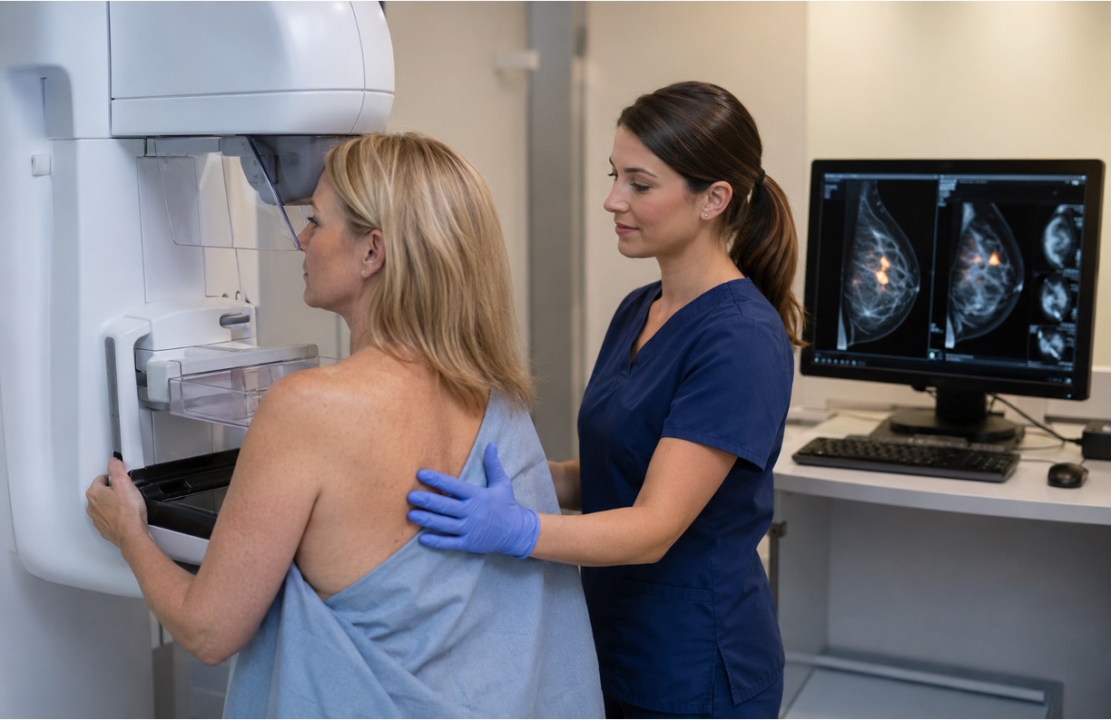
- The FDA cleared Siemens Healthineers' breast imaging system for both contrast-enhanced mammography (CEM) and CEM-guided biopsy, allowing centers to detect and sample enhancing lesions on a single platform.
- CEM biopsy follows a stereotactic technique with added iodinated contrast injection and dual-energy imaging, all performed within the contrast's limited enhancement window.
- Dense breast tissue masks lesions on standard mammography, but CEM shifts detection to vascularity-based contrast, making enhancing malignancies visible regardless of fibroglandular density.
- CEM biopsy also serves patients who cannot undergo MRI due to pacemakers, implants, claustrophobia, or renal limits, plus cases where a lesion enhances on CEM but has no MRI correlate.
- ContrastConnect provides immediate, CMS-compliant virtual contrast supervision for CEM workflows, with audit-ready documentation and radiologists who manage over 130 contrast reactions monthly.
The Siemens Mammomat B.brilliant FDA 510(k) Clearance
Siemens Healthineers received FDA clearance for a breast imaging system that can perform both contrast-enhanced mammography (CEM) and CEM-guided biopsy on one machine. In practice, that means a facility can spot a suspicious lesion and take a tissue sample without moving the patient to a different suite or switching to a different imaging method.
The clearance covers the full workflow: acquiring contrast-enhanced images and then using those same images to guide a biopsy needle to the target. Before this, the two steps often required separate equipment or departments.
The system also includes ClearCEM, Siemens' proprietary image reconstruction technology. Standard CEM processing can produce artifacts that blur lesion edges or reduce image clarity. ClearCEM applies advanced reconstruction algorithms to clean up those subtracted images, producing sharper views of enhancing lesion boundaries, which matters most when those images are guiding a needle to an exact location.
Because every CEM biopsy begins with an iodinated contrast injection, ContrastConnect provides the real-time virtual supervision and audit-ready documentation facilities need to keep each case CMS-compliant from injection through sampling.

ContrastConnect: Virtual Contrast Supervision That Never Misses
Built by Radiologists | 75,000+ Hours Monthly Contrast Exams | Trusted Nationwide

Built for Imaging Networks:
- Virtual Contrast Supervision: Radiologists provide immediate CMS-compliant supervision through a secure, HIPAA-compliant platform for outpatient facilities and hospital networks.
- Unmatched Experience: 130+ contrast reactions treated monthly with 3,900+ technologists certified.
The ContrastConnect Difference:
- ✓ Radiologist-owned with superior clinical expertise
- ✓ Always-on platform with guaranteed compliance
- ✓ Audit-ready documentation for CMS reviews
- ✓ Cost-efficient alternative to onsite staffing
Safety & Compliance You Trust:
Helping imaging centers reduce cancellations, extend hours, and scale operations without adding on-site radiologists. Response times measured in seconds.
CEM Biopsy Workflow: Step-by-Step
CEM-guided biopsy follows a structured sequence that borrows heavily from stereotactic biopsy technique but integrates contrast administration and dual-energy image acquisition as foundational steps. The workflow is more time-sensitive than a standard stereotactic procedure because iodinated contrast has a finite enhancement window, and the biopsy targeting must occur while the lesion remains visible on recombined images.
- Patient Preparation and Contrast Injection: Before the procedure begins, patients must be screened for iodinated contrast contraindications, including renal insufficiency, prior contrast reactions, and metformin use. Informed consent should address both the biopsy and the contrast administration. IV access is established, typically in the antecubital fossa, and the contrast agent is injected at a standard weight-based dose prior to image acquisition.
- Image Acquisition and Lesion Targeting: Once contrast is administered and the breast is positioned in compression, dual-energy image pairs are acquired. The low-energy images resemble a conventional mammogram, while the high-energy images capture differential contrast uptake. The radiologist reviews the recombined views to confirm lesion visibility and conspicuity before proceeding to coordinate-based targeting.
- Scout Image Use in Tomosynthesis-Guided Biopsy: The tomosynthesis scout image serves as the procedural bridge between contrast-enhanced lesion identification and physical needle placement. Rather than relying on 2D projections alone to estimate depth, the tomosynthesis scout provides a three-dimensional reference that allows the system to calculate a precise Z-axis coordinate for the needle.
- Sampling Technique and Post-Procedure Protocol: Core needle biopsy is performed using standard vacuum-assisted technique, with needle gauge selected based on lesion size and location. Real-time imaging during sampling confirms the needle remains on-target within the enhancing zone. A biopsy marker clip is then deployed at the sampling site immediately following tissue acquisition.

Clinical Use Cases for CEM-Guided Biopsy
Dense Breast Tissue Evaluation
Dense breast tissue is the most common driver of diagnostic difficulty in mammography. Fibroglandular tissue and malignant lesions share similar radiographic density on standard 2D mammography, masking lesions that would be visible on contrast-enhanced imaging.
CEM cuts through this problem by shifting lesion detection from density-based contrast to vascularity-based contrast. An enhancing malignancy stands out on recombined CEM images regardless of background fibroglandular density.
For women with heterogeneously dense or extremely dense breasts who have a suspicious finding on an adjunct imaging modality but no ultrasound correlate, CEM-guided biopsy provides a direct sampling path without the cost, time, and access barriers associated with MRI-guided biopsy.
Enhancing Lesions With No MRI Correlate
While MRI remains one of the best options for breast soft tissue evaluation, discordance between CEM and MRI findings does occur. And when a lesion enhances on CEM but lacks a clear MRI correlate, CEM-guided biopsy becomes the only viable direct sampling route.
This scenario is more common than it might seem, particularly for lesions near the chest wall, in the axillary tail, or in areas where MRI artifact or patient motion degrades image quality. CEM-guided biopsy ensures that enhancing lesions found on CEM don't go unsampled simply because MRI guidance isn't feasible or concordant.
Patients Who Cannot Undergo MRI
Patients with pacemakers, certain implanted devices, severe claustrophobia, or compromised renal function that prevents gadolinium administration represent a substantial subset of women who need diagnostic workup but cannot safely undergo breast MRI.
For these patients, CEM has historically offered better lesion detection than standard mammography alone. But without a biopsy pathway, acting on CEM findings required either deferring to less sensitive guidance or waiting for an alternative approach.
CEM-guided biopsy closes that loop. A patient who cannot tolerate MRI but has a suspicious enhancing lesion on CEM now has a direct, image-concordant biopsy option using the same modality that identified the lesion.
This is clinically meaningful because it eliminates the diagnostic dead end that previously existed for this group and aligns the detection and sampling workflow within a single imaging platform.
Keep Your CEM Program Compliant & Audit-Ready With ContrastConnect
CEM-guided biopsy is a meaningful step forward for breast imaging, giving centers a way to detect and sample enhancing lesions on one platform and reaching patients who were previously stuck at a diagnostic dead end. Because a CEM biopsy begins with an iodinated contrast injection, it falls under the same CMS direct-supervision requirement as any contrast study, and it carries real reaction risk during a time-sensitive procedure.
At ContrastConnect, we deliver immediate, CMS-compliant virtual supervision through a secure platform, supported by experienced radiologists who manage 130+ contrast reactions each month and produce audit-ready documentation built for CMS review. As you build or expand a CEM biopsy service, talk to us about keeping every case supervised, safe, and ready for an audit. Start your coverage assessment now.
Frequently Asked Questions (FAQs)
Is contrast-enhanced mammogram FDA approved?
CEM is not currently FDA approved for screening use. However, diagnostic use of CEM as an alternative to breast MRI is widely accepted and growing in clinical practice. The FDA has cleared specific CEM and CEM biopsy features, including those on the Siemens Healthineers breast imaging platform, for diagnostic and procedural applications.
What are the main clinical applications of mammography?
Standard mammography is primarily used for breast cancer screening and diagnostic workup of palpable or symptomatic breast findings. In screening, it detects suspicious masses, calcifications, architectural distortions, and asymmetries that may indicate early malignancy. Diagnostic mammography adds targeted views and spot compression to characterize findings identified on screening.
When did contrast-enhanced mammography start?
CEM was first described in the medical literature by Watt et al. in 1985 using digital subtraction angiography of the breast, though the dual-energy subtraction technique used in modern CEM systems was first described by Lewin et al. in 2003. Clinical adoption accelerated after the FDA approved CEM for diagnostic use in 2011.
How does CEM biopsy workflow differ from MRI-guided biopsy?
CEM-guided biopsy and MRI-guided biopsy both target enhancing breast lesions, but the procedural mechanics, time requirements, and logistical demands differ considerably. MRI-guided biopsy requires a dedicated MRI suite, MRI-compatible biopsy equipment, and a contrast agent that carries its own contraindication profile. The procedure typically takes longer and is resource-intensive in terms of scheduling and equipment use.
Does CMS recognize remote supervision as direct supervision?
Yes. Effective January 1, 2026, CMS permanently updated its definition of direct supervision so the supervising physician can meet the immediate-availability requirement through real-time, two-way audio and video technology instead of being on-site. At ContrastConnect, we connect your facility to an experienced radiologist in real time so every contrast study stays supervised, compliant, and fully documented for audits.
*Note: Information provided is for general guidance only and does not constitute medical, legal, or financial advice. Pricing estimates and regulatory requirements are current at the time of writing and subject to change. For personalized consultation on imaging center operations and virtual contrast supervision, contact ContrastConnect.
Trusted Nationwide














.avif)




1,000,000
Contrast exams supervised annually
75,000+
Hours of supervision monthly
3,900+
Technologists certified
100s
Of imaging partners nationwide
130+
Contrast reactions treated monthly
100%
Requested hours covered
Connect with us.

